One-Pager
Electrolyte Imbalances
Na, K, Ca, Mg and phos: normals, ECG clues, safe replacement, hyperkalemia steps
Download one-page PDFLetter landscape, ready to print or keep with your notes
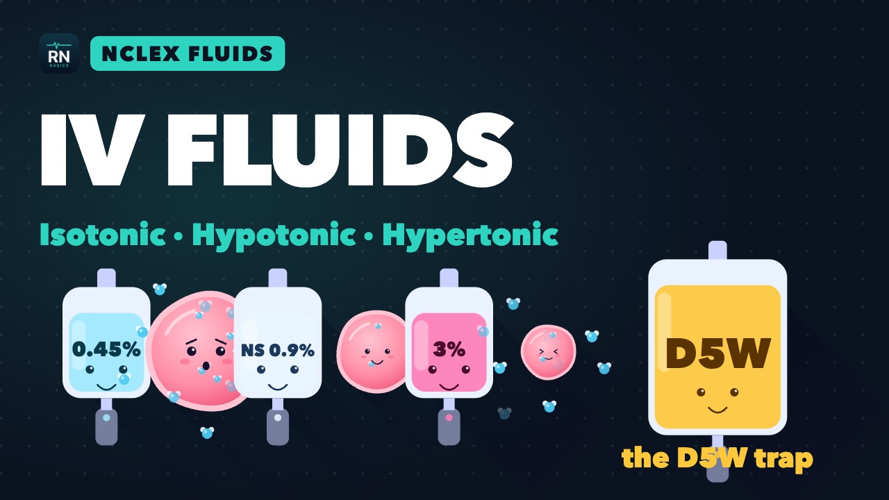
Watch the explainer
IV Fluids Explained: Isotonic, Hypotonic, Hypertonic (NCLEX)
Normals and key signs
Exam study summary, not a bedside tool. Doses are typical adult ranges; follow your institution's protocols.
| Ion | Normal | Low | High |
|---|---|---|---|
| Na | 135–145 mEq/L | Nausea, confusion, seizures | Thirst, confusion, seizures |
| K | 3.5–5.0 mEq/L | Weakness; flat T, U wave, ST sag | Weakness; peaked T, wide QRS |
| Ca | 8.8–10.3 mg/dL; ionized 4.7–5.2 mg/dL (1.17–1.30 mmol/L) | Tetany, Chvostek, Trousseau; long QT | Stones, constipation, confusion; short QT |
| Mg | 1.7–2.2 mg/dL | Tremor, tetany; long QT, torsades | Lost reflexes, slow breathing, long PR |
| Phos | 2.8–4.5 mg/dL | Weakness, failure to wean, hemolysis | Low-Ca signs (tetany, long QT) |
Hyperkalemia: stabilize, shift, remove
- Stabilize (ECG changes): calcium gluconate 10% 30 mL over 10 min; in arrest or peri-arrest, calcium chloride 10% 10 mL over 5 min (same calcium). Acts in about 3 min, lasts 30–60 min; repeat if changes persist.
- Shift: regular insulin 10 units IV with dextrose 25–50 g. K falls within 15 min, lowest at 30–60 min.
- Add-on shift: nebulized albuterol 10–20 mg, never alone. Bicarbonate mainly for metabolic acidosis.
- Remove: binders (sodium zirconium cyclosilicate acts in about 1 h, patiromer 4–7 h), loop diuretic, dialysis (definitive).
- Recheck K often; it rebounds as the shift wears off.
In cardiac arrest (AHA 2025), calcium, bicarbonate and insulin-glucose may be considered but are unproven; inhaled albuterol is not recommended.
Potassium and magnesium replacement
- Check urine output and kidney function first.
- IV KCl: always diluted, always on a pump.
- Peripheral IV: usually no faster than 10 mEq/h.
- Central line, continuous ECG: often up to 20 mEq/h per policy; the label allows 40 only if K is under 2 with ECG changes or paralysis.
- DKA: if K is below 3.5, give K first and hold insulin (ADA 2024).
- Torsades (polymorphic VT with long QT): magnesium sulfate 1–2 g IV; defibrillate (unsynchronized) if pulseless or sustained. Mg is not advised for normal-QT polymorphic VT.
- Mg toxicity (preeclampsia drip): reflexes fade, then breathing slows. Stop the Mg; give IV calcium gluconate.
Calcium: salts and corrected calcium
| Feature | Gluconate 10% | Chloride 10% |
|---|---|---|
| Elemental Ca per 10 mL | About 93 mg | About 270 mg (3 times more) |
| Line | Peripheral OK (secure IV) | Central or deep vein; necrosis if it leaks |
Corrected Ca = Ca + 0.8 × (4.0 − albumin g/dL). Example: Ca 7.6, albumin 2.0 → 9.2 mg/dL, not truly low.
Low ionized Ca: alkalosis, massive transfusion (citrate), thyroid or parathyroid surgery, pancreatitis.
High Ca: IV saline first. If severe (over 14 mg/dL), calcitonin (fast, fades in 48–72 h) plus an IV bisphosphonate or denosumab (slower, longer acting).
Sodium: correct slowly
- Severe symptoms (seizures, coma): 3% saline 100–150 mL over 10–20 min, repeated to raise Na about 5 mEq/L.
- Chronic: raise Na no more than 8 mEq/L in any 24 h; guideline upper limits run 10–12.
- ODS risk: very low Na, alcohol use, malnutrition, liver disease, low K.
- ODS shows days later: dysarthria, dysphagia, paralysis.
- Overcorrected: stop saline; re-lower with D5W, desmopressin on expert advice.
- Hypernatremia: give free water; lower chronic cases slowly, over about 48 h.
Causes, refeeding, phosphate
- High K: AKI or CKD, ACE inhibitors, ARBs, spironolactone, rhabdo or burns, acidosis, transfusion.
- Tumor lysis: high K, phos and uric acid; low Ca.
- Refeeding: phos, K and Mg fall within 5 days of restarting nutrition (ASPEN 2020).
- IV phosphate: slowly over hours, never with calcium in the same line.
In depth
The one-page summary above is what the PDF prints. These sections go further.
Sodium, water and tonicity
Serum Na measures water balance, not body sodium: Na ≈ (exchangeable Na + K) ÷ total body water (Edelman). Low Na almost always means ADH is stopping the kidney from excreting water; high Na means water losses outran intake in someone who cannot drink. Because K is in the numerator, KCl raises Na much as NaCl does.
Tonicity (effective osmolality) = 2 × Na + glucose ÷ 18; urea and ethanol cross cell membranes, so they raise measured osmolality without moving water. ADH release starts near 280 mOsm/kg and thirst near 290 (European guideline 2014). A 5% fall in blood pressure also raises ADH, as do nausea, pain, anesthesia and many drugs.
Water excretion is limited by solute: maximum urine volume ≈ solute intake ÷ minimum urine osmolality (about 50 mOsm/kg). A beer or tea-and-toast diet of 250 mOsm/day allows only 5 L of urine, so drinking more lowers Na even without ADH.
Over 24–48 h, brain cells shed electrolytes and organic osmolytes to undo swelling (European guideline 2014). That sets the 48 h line between acute and chronic hyponatremia and makes fast correction dangerous: osmolytes return slowly, so a quick rise shrinks adapted cells and strips myelin. In hypernatremia the risk is reversed: cerebral edema.
Hyponatremia: work-up by urine and volume
Send urine osmolality and Na with the serum sample, before saline or diuretics change them. The European guideline (2014) works in this order: serum osmolality under 275 mOsm/kg confirms hypotonicity; urine osmolality 100 or less means intake exceeds maximal excretion (polydipsia, beer potomania); above 100, urine Na 30 mEq/L or less means low effective arterial volume. Volume status comes last because bedside assessment is unreliable.
| Volume | Urine Na | Causes | First step |
|---|---|---|---|
| Low, extrarenal | Under 30 | Diarrhea, vomiting, burns, third spacing | Isotonic saline or balanced fluid |
| Low, renal | Over 30 | Thiazides, salt wasting, adrenal insufficiency | Saline; stop the drug; hydrocortisone |
| Normal | Over 30 | SIADH, hypothyroidism, low cortisol | Restrict water; treat the cause |
| Normal, dilute urine | Often low | Polydipsia, low solute intake | Stop excess water; add solute |
| High | Under 30 off diuretics | Heart failure, cirrhosis, nephrotic syndrome | Restrict water; loop diuretic |
SIADH is a diagnosis of exclusion: effective osmolality under 275, urine osmolality over 100, euvolemia, urine Na over 30, normal adrenal, thyroid and kidney function, and no recent diuretic; uric acid under 4 mg/dL supports it (European guideline 2014). ICU causes: pneumonia, positive-pressure ventilation, SAH, head trauma, pain, nausea, SSRIs, carbamazepine, opioids, cyclophosphamide, vasopressin analogues.
Cerebral salt wasting shares the labs but adds volume depletion and high urine output; it needs salt and volume. After SAH, fluid restriction raises the risk of delayed ischemia, so it is avoided even when SIADH is likely. In SIADH, 0.9% saline lowers Na further when urine Na + K exceeds 154 mEq/L: the salt leaves in less water than it came in with.
Hyponatremia: correction limits and overcorrection rescue
Severe symptoms (vomiting, seizures, deep somnolence, coma) need 3% saline now, acute or chronic: Europe 2014 gives 150 mL over 20 min, repeated up to twice until Na has risen 5 mEq/L; Merck gives 100 mL over 15 min. In SALSA (2021, 178 patients), boluses reached target sooner than slow infusion without more overcorrection. Then stop 3% and treat the cause.
| Source | Limit (mEq/L) | High ODS risk |
|---|---|---|
| Europe 2014 | 10 in the first 24 h, then 8 per 24 h | No separate limit |
| US panel 2013 | 10–12 in any 24 h; 18 in any 48 h | 8 in any 24 h |
| Merck 2025 | 8 in the first 24 h | No separate limit |
High ODS risk (US panel 2013): Na 105 or less, hypokalemia, alcohol use disorder, malnutrition, liver disease. In a 2025 meta-analysis (Kidney Med; 26,710 patients), ODS occurred in 0.23% overall and 2.9% with Na under 116; correction over 8 mEq/L in 24 h tripled the odds, yet about a third of cases had no rapid correction.
Overcorrection usually comes from the patient: once the ADH stimulus ends (volume restored, thiazide stopped, cortisol given, nausea relieved), dilute urine pours out. With Na under 120, the rise beat the Adrogué–Madias formula (next section) in 74% of patients, by 2.4 times on average (Europe 2014). Rising urine output with falling specific gravity means check Na now.
- Trigger (Europe 2014): Na up more than 10 in the first 24 h or 8 in any later 24 h; stop all sodium-raising treatment.
- With expert advice, give D5W 10 mL/kg over 1 h, with strict intake and output.
- Add desmopressin 2 mcg IV, no more often than every 8 h, to stop the water diuresis.
The desmopressin clamp gives desmopressin 1–2 mcg IV or SC every 6–8 h with slow 3% saline and no free water, so Na rises only with the sodium given (Sood 2013: 5.8 mEq/L in 24 h, no overcorrection). Evidence is mixed: proactive use cut overcorrection from 27.1% to 6.8% in a 2025 cohort (AlShanableh), but a 49-patient trial found no significant difference (Pakchotanon 2024). Any free water on desmopressin drops Na.
Chronic SIADH: restrict fluid first; restriction cannot work when urine Na + K exceeds serum Na (Furst 2000). Second line (Europe 2014): urea 0.25–0.50 g/kg/day or a loop diuretic with oral salt. Tolvaptan starts in hospital, with no fluid restriction for 24 h, for at most 30 days (label: liver injury); Europe advises against vaptans. Hypovolemia: 0.9% saline or balanced fluid at 0.5–1 mL/kg/h (Europe 2014).
Hypernatremia and the free water deficit
Hypernatremia is always hypertonic and nearly always a water deficit in someone who cannot drink. ICU causes: fever, osmotic diuresis (glucose, mannitol, high-protein feeds), loop diuretics, lactulose and sodium loads (bicarbonate, hypertonic saline).
- Small volume of concentrated urine: loss outside the kidney, or no intake.
- 3–30 L/day of dilute urine (SG under 1.005): diabetes insipidus; a rise in urine osmolality of 50% or more after desmopressin means central DI (Merck).
- Central DI: pituitary surgery, head trauma, SAH, brain death. Nephrogenic: lithium, high Ca, low K.
Free water deficit = TBW × (Na ÷ 140 − 1), with TBW 0.6 × weight in younger men, 0.5 in younger women and older men, 0.45 in older women (Europe 2014). An 80 kg man with Na 162: TBW 48 L, deficit about 7.5 L. Lowering Na by 10 in a day takes 48 × (162 ÷ 152 − 1) ≈ 3.2 L plus ongoing losses. Adrogué–Madias gives the change per liter as (infusate Na + K − serum Na) ÷ (TBW + 1): D5W lowers his Na about 3.3 mEq/L per liter (162 ÷ 49), 0.45% saline about 1.7.
Merck lowers chronic or unknown-duration hypernatremia over 48 h at no more than 0.5 mEq/L/h; Adrogué and Madias (2000) target 10 mEq/L per day, faster if the rise took hours. In Chauhan 2019 (449 ICU patients, Na over 155), the 154 corrected faster than that had no excess mortality or cerebral edema. Exams still expect slow correction.
Give water enterally when possible; D5W is pure free water (watch glucose) and 0.45% saline is half. In shock, restore perfusion first: 0.9% saline is still hypotonic to a Na of 160. Treat the cause: desmopressin for central DI; stop lithium and correct Ca and K for nephrogenic DI.
Potassium: shifts, losses and replacement
A 70 kg adult holds about 3,500 mEq of K, 98% inside cells (Merck). Insulin, beta-2 agonists and alkalosis drive K in; hyperosmolality, normal-gap acidosis, cell lysis, digoxin toxicity and succinylcholine move it out, while lactic and ketoacidosis shift little (Merck). Secretion in the collecting duct needs aldosterone, distal sodium delivery and urine flow, so ACE inhibitors, spironolactone, trimethoprim, heparin and oliguria raise K, and loop or thiazide diuretics and vomiting lower it.
Each 1 mEq/L fall reflects a loss of about 200–400 mEq (Merck), so a K of 2.5 can need 300–600 mEq over days. Shift hypokalemia (insulin, beta agonists, hypothermia, thyrotoxic periodic paralysis) is the exception: replacement overshoots when the shift reverses, as on rewarming after cooling. Urine K over 15 mEq/L points to renal loss (Merck).
- Oral when the gut works and the ECG is normal: 20–80 mEq/day in divided doses (Merck).
- KCl when chloride is low (vomiting, diuretics); K phosphate when phosphate is low too; acetate or citrate in acidosis.
- Replace Mg at the same time; K will not hold while Mg is low.
- DKA (ADA 2024): add 20–30 mEq per liter once K is under 5.0, aiming for 4–5; hold insulin until K is above 3.5.
UKKA 2026 grades high K as mild (5.5–5.9), moderate (6.0–6.4) or severe (6.5 or more) and monitors severe cases continuously. Insulin 10 units with 25 g glucose lowers K by 0.65–1.4 mEq/L, and up to 40% of patients with end-stage kidney disease do not respond to salbutamol. Besides hemolysis, pseudohyperkalemia comes from fist clenching, thrombocytosis and extreme leukocytosis (Merck); a blood gas K is a quick check.
Calcium: ionized vs total, PTH, citrate and malignancy
About 40% of serum Ca is protein-bound, mostly to albumin, about half is ionized and the rest complexed (Merck); only ionized Ca acts. Alkalosis binds Ca to albumin, so hyperventilation or bicarbonate lowers iCa with total Ca unchanged. In Özdemir 2026 (250 ICU patients), albumin-corrected Ca caught only 15.2% of low iCa, uncorrected total Ca 62.8%.
A fall in iCa raises PTH within minutes: Ca leaves bone, the kidney keeps Ca and wastes phosphate, and activated vitamin D raises gut absorption over days. Mg under about 1.0 mg/dL blocks PTH release and action (Merck), so replace Mg first. Beyond the one-pager's list, low iCa follows sepsis, CRRT citrate, tumor lysis and rhabdomyolysis.
The liver clears citrate in minutes, but shock, hypothermia and liver failure slow it, so iCa falls during large transfusions (Merck: over 10 units): measure iCa often and keep it normal. For symptoms, give calcium gluconate 10% 10 mL over 10 min, then 20–30 mL in 1 L D5W over 12–24 h (Merck). Gluconate needs no liver: both salts raised iCa equally in anhepatic patients (Martin 1990). Treating mild ionized hypocalcemia has no proven benefit (Cochrane, Forsythe 2008), so thresholds vary.
High Ca comes mostly from hyperparathyroidism (PTH high or normal) or cancer (PTH suppressed). In cancer, PTH-related peptide causes about 80%, bone metastases or myeloma about 20%, and lymphoma calcitriol under 1% (Stewart 2005). It blocks urine concentration, so patients arrive dry: fluid comes first. Severe is over 14 mg/dL (Endocrine Society 2023).
| Treatment | Dose | Onset | Notes |
|---|---|---|---|
| Isotonic saline | 1–2 L over 2–4 h, then urine about 250 mL/h (Merck) | Hours | Tailor to cardiac function |
| Calcitonin | 4–8 units/kg IM or SC every 6–12 h | 4–6 h; Ca falls 1–2 mg/dL | Tachyphylaxis; limit to 48–72 h |
| Zoledronic acid | 4 mg IV over at least 15 min (label) | 48–72 h | No cut for creatinine under 4.5 (label); check it each dose |
| Denosumab | 120 mg SC | 3–10 days | Hypocalcemia risk if GFR under 30 |
| Glucocorticoids | Prednisone 40–60 mg/day (Merck) | Days | Calcitriol-driven: lymphoma, granulomas |
| Hemodialysis | Low-calcium dialysate | Hours | Ca over 18 mg/dL or severe symptoms (Merck) |
Magnesium, phosphate and refeeding
About 1% of body Mg is in serum, so a normal level does not exclude depletion. Causes: loop and thiazide diuretics, PPIs, alcohol, diarrhea, amphotericin, cisplatin, calcineurin inhibitors, refeeding and CRRT. Low cell Mg releases its brake on ROMK channels, so the distal nephron wastes K until Mg is restored (Huang 2007).
Merck gives magnesium sulfate 2–4 g IV over 5–10 min for seizures or arrhythmia (AHA uses 1–2 g for torsades), otherwise about 1 g/h for up to 10 h, because about half of an IV dose is lost in urine; give less in kidney failure. High Mg (Merck): ECG changes at 6–12 mg/dL, reflexes lost near 12, then hypotension and respiratory depression, arrest above 15. Give calcium gluconate 10% 10–20 mL IV, and dialysis if the kidneys have failed.
Phosphate builds ATP and 2,3-DPG. Low levels (severe under 1 mg/dL, Merck) come from shifts (refeeding, insulin, respiratory alkalosis), CRRT losses, burns and alcohol use disorder. Effects: diaphragm weakness and failure to wean, poor contractility, rhabdomyolysis, hemolysis and a left-shifted oxyhemoglobin curve. In Aubier 1985, 8 ventilated patients generated more diaphragm pressure after 10 mmol of phosphate over 4 h.
| Serum phos (mg/dL) | IV phosphate dose | K delivered |
|---|---|---|
| 1.8 to low normal | 0.16–0.31 mmol/kg | 0.23–0.46 mEq/kg |
| 1.0–1.7 | 0.32–0.43 mmol/kg | 0.47–0.63 mEq/kg |
| Under 1.0 | 0.44–0.64 mmol/kg | 0.64–0.94 mEq/kg |
Normalize Ca first and never run phosphate with calcium (label). In kidney failure, tumor lysis or rhabdomyolysis, high phosphate binds Ca; precipitation is likely once Ca × phos exceeds about 70 (Merck).
Refeeding syndrome (ASPEN 2020) is a fall in phos, K or Mg of 10–20% (mild), 20–30% (moderate), or over 30% or with organ dysfunction (severe) within 5 days of restarting calories. High risk includes BMI under 16 or little intake for over 7 days.
- Check K, Mg and phos before feeding, then every 12 h for 3 days if high risk.
- Start with 100–150 g of dextrose or 10–20 kcal/kg for 24 h; advance by a third of goal every 1–2 days.
- Thiamine 100 mg before feeding or dextrose, then daily for 5–7 days in high risk.
Electrolytes on the ECG
| Disturbance | ECG | Rhythm risk |
|---|---|---|
| K over 5.5 | Tall peaked T; PR lengthens | Rises with level and speed of rise |
| K over 6.5 | Wide QRS; P waves fade | Junctional and escape rhythms, bradycardia |
| Severe high K | Sine wave | VF, asystole; pacing may fail |
| Low K | Flat T, ST sag, U wave, long QU | PVCs, VT, torsades; digoxin toxicity |
| Low Mg | Long QT | Torsades; digoxin toxicity |
| Low Ca | Long QT from a long ST segment | Torsades is rare |
| High Ca | Short QT (short ST segment) | Bradycardia, AV block |
| High Mg 6–12 mg/dL | Long PR, wide QRS, taller T | Heart block; arrest over 15 mg/dL |
The ECG rules high K in, not out: in the study UKKA 2026 cites, sensitivity was 0.19 and specificity 0.97, and the first sign can be VF or asystole. QRS widening, heart rate under 50 and junctional rhythm predict adverse events; that bradycardia may resist atropine and pacing, pacemakers can malfunction above about 7 mEq/L, and IV calcium can raise the rate (UKKA 2026).
Low K, low Mg and QT-prolonging drugs add up to torsades. Repeat the ECG after each corrective step.
Dialysis, CRRT and safe replacement
Hemodialysis removes 70–100 mEq of K per session; serum K falls about 1 mEq/L in the first hour and 1 more over the next 2 hours, then rebounds as cells release K, more after insulin or salbutamol (UKKA 2026). Give IV calcium for ECG changes even when dialysis is about to start. CRRT lowers K more slowly but with less rebound; K follows the bag (0, 2 or 4 mEq/L).
On CRRT, phosphate-free fluid causes hypophosphatemia in most patients by day 2–3, Mg is lost (more on citrate), and citrate accumulation shows as falling iCa with rising total Ca, a total-to-ionized ratio of 2.5 or more (CRRT sheet). Dialysis pulls Na toward the fluid's 140, so chronic hyponatremia can overcorrect within hours; units lower the fluid Na, by methods that vary.
- Concentrated KCl stays in pharmacy (Joint Commission 2003); hang premixed bags on a pump. Never push K or add it to a hanging bag, where it can layer (label).
- KCl label: up to 10 mEq/h and 200 mEq/day if K is over 2.5; 40 mEq/h and 400 mEq/day only for K under 2 with ECG changes or paralysis, on continuous ECG.
- Labels allow 40 mEq/L peripherally and keep 300–400 mEq/L central; many hospitals still run 10 mEq/100 mL peripherally and 20 mEq/h centrally. Follow policy.
- Continuous ECG above 10 mEq/h (K phosphate label). Check urine output first; count K phosphate, PN, feeds and CRRT fluid.
- High-alert (ISMP): concentrated KCl, K phosphate, hypertonic saline and IV Mg get an independent double check and smart-pump limits.
- Calcium chloride goes central (necrosis if it leaks); never with bicarbonate, phosphate or ceftriaxone in the line.
- 3% saline runs peripherally in many protocols, 7.5% and stronger centrally (ENLS); check Na every 4 h on 3% (Europe 2014).
Exam pearls
- SIADH is euvolemic (urine osmolality over 100, urine Na over 30): restrict fluid, but not after SAH. Cerebral salt wasting is volume depleted: give saline.
- DI: lots of dilute urine (SG under 1.005) and rising Na; central DI responds to desmopressin.
- Chronic hyponatremia: no more than 8 mEq/L in 24 h; reverse overcorrection with D5W and desmopressin.
- Free water deficit = TBW × (Na ÷ 140 − 1); lower chronic hypernatremia about 10 mEq/L a day.
- High K: peaked T, long PR and lost P, wide QRS, sine wave; calcium first for ECG changes.
- Low K: flat T, U waves; replace Mg too; never push KCl.
- Low Ca: Chvostek, Trousseau, long QT after neck surgery, transfusion or pancreatitis; check ionized Ca.
- High Ca: short QT, polyuria, confusion; saline, then calcitonin plus a bisphosphonate or denosumab.
- High Mg: reflexes fade before breathing slows; stop it and give calcium gluconate.
- Low phos: weakness and failure to wean after refeeding; thiamine before feeding.
Related one-pagers
- Sepsis and Septic Shock BundleSepsis-3 definitions, the Hour-1 bundle and SSC 2026 changes for exam review
- DKA and HHS2024 consensus criteria, treatment order, potassium, euglycemic DKA and monitoring
- Blood Products and TransfusionComponents, triggers, compatibility, reactions, massive transfusion, reversal
- Acute Abdomen and GI Surgical EmergenciesObstruction and NG decompression, ileus and Ogilvie, mesenteric ischemia, perforation, Whipple and bariatric leaks, dumping and Fournier gangrene
- Lab Values to KnowNormal ranges in US units, therapeutic drug levels, and acting on a critical result
Calculators for this topic
Sources
- Alfonzo A, Harrison A, Baines R, Chu A, Mann S, MacRury M. Clinical Practice Guidelines: Treatment of Acute Hyperkalaemia in Adults. UK Kidney Association; original July 2020, updated October 2023, updated July 2026 (review date October 2026). Link
- Part 10: Adult and Pediatric Special Circumstances of Resuscitation: 2025 American Heart Association Guidelines for CPR and ECC. Circulation. 2025. doi:10.1161/CIR.0000000000001380 Link
- Part 9: Adult Advanced Life Support: 2025 American Heart Association Guidelines for CPR and ECC. Circulation. 2025. doi:10.1161/CIR.0000000000001376 Link
- Neumar RW, et al. Part 8: Adult Advanced Cardiovascular Life Support: 2010 AHA Guidelines for CPR and ECC. Circulation. 2010;122:S729-S767. Link
- Merck Manual Professional Version: Long QT Syndrome and Torsades de Pointes Ventricular Tachycardia (reviewed September 2024). Link
- Umpierrez GE, Davis GM, ElSayed NA, et al. Hyperglycemic Crises in Adults With Diabetes: A Consensus Report. Diabetes Care. 2024;47:1257. doi:10.2337/dci24-0032 Link
- Spasovski G, Vanholder R, Allolio B, et al. Clinical practice guideline on diagnosis and treatment of hyponatraemia. Eur J Endocrinol. 2014;170(3):G1-G47. Link
- Hyponatremia Correction and Osmotic Demyelination Syndrome Risk: A Systematic Review and Meta-Analysis. Kidney Med. 2025. doi:10.1016/j.xkme.2024.100953 Link
- Potassium Chloride for Injection Concentrate, prescribing information (Hospira). DailyMed, US National Library of Medicine. Link
- Potassium Chloride Injection (premixed), prescribing information (ICU Medical). DailyMed. Link
- Calcium Chloride Injection 10%, prescribing information (Hospira). DailyMed. Link
- Calcium Gluconate Injection, prescribing information (WG Critical Care). DailyMed. Link
- Merck Manual Professional Version: Hypokalemia; Hyponatremia; Hypernatremia (Electrolyte Disorders). Link
- Merck Manual Professional Version: Hyperkalemia. Link
- Merck Manual Professional Version: Hypocalcemia; Hypercalcemia. Link
- Merck Manual Professional Version: Hypomagnesemia; Hypermagnesemia. Link
- Merck Manual Professional Version: Hypophosphatemia; Hyperphosphatemia. Link
- Merck Manual Professional Version: Preeclampsia and Eclampsia. Link
- Merck Manual Professional Version: Management of Adverse Effects of Cancer Therapy (tumor lysis syndrome). Link
- MedlinePlus Medical Encyclopedia: Calcium blood test (reviewed 11/6/2025), Magnesium blood test (reviewed 2/13/2025) and Phosphorus blood test (reviewed 11/6/2025). US National Library of Medicine. Link
- MedlinePlus Medical Encyclopedia: Sodium blood test. US National Library of Medicine. Link
- El-Hajj Fuleihan G, et al. Treatment of Hypercalcemia of Malignancy in Adults: An Endocrine Society Clinical Practice Guideline. J Clin Endocrinol Metab. 2023;108(3):507-528. Link
- da Silva JSV, et al. ASPEN Consensus Recommendations for Refeeding Syndrome. Nutr Clin Pract. 2020;35(2):178-195. Link
- Özdemir E, Yılmaz T, Düzenci D. Limited diagnostic utility of albumin-corrected calcium in the intensive care unit: a prospective comparison with ionised calcium. PLOS One. 2026;21(7):e0354233. doi:10.1371/journal.pone.0354233 Link