 RN Basics
Video explainer · Rhythms
EKG Rhythms Explained: AFib, Flutter, Heart Blocks, VT, VF and Asystole
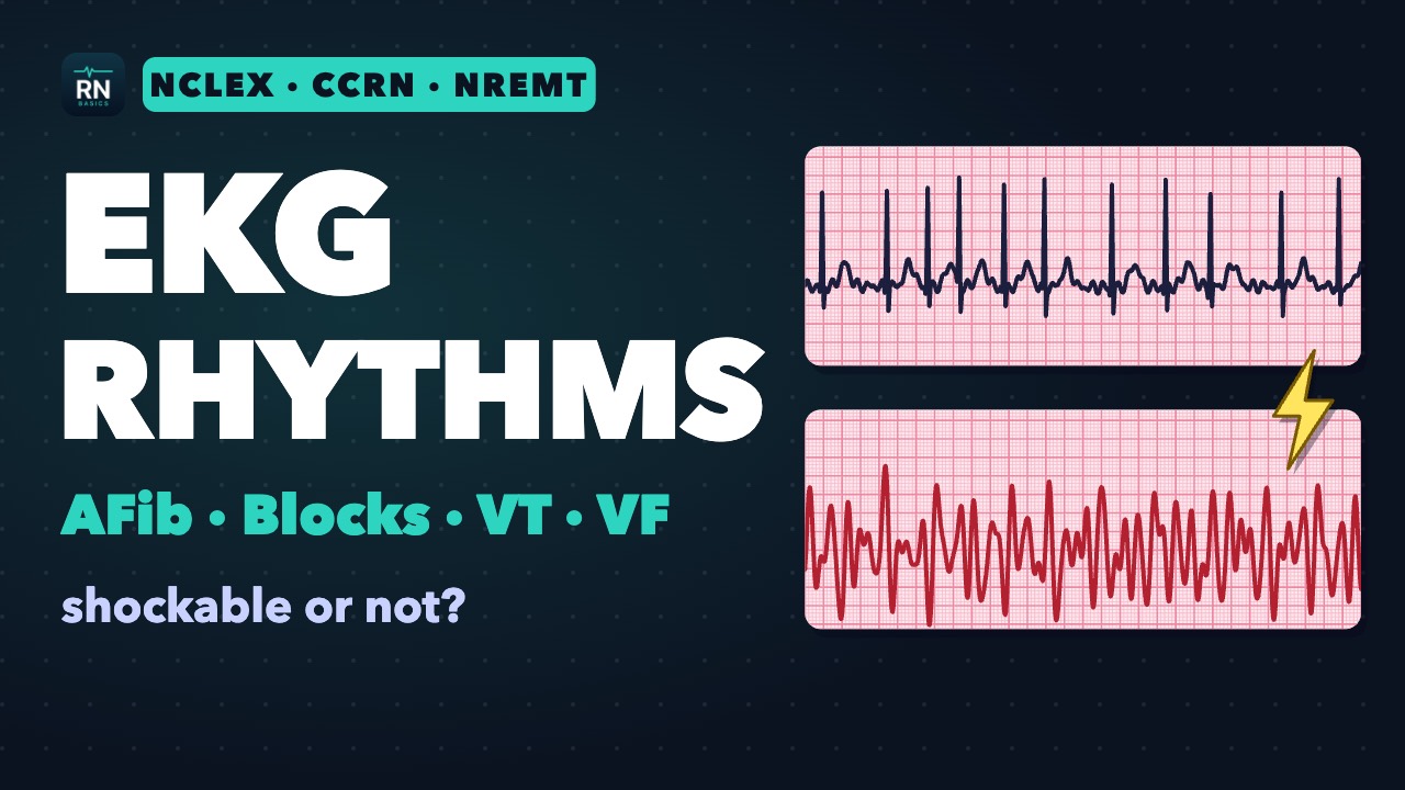
How to read a rhythm strip in five steps, then the rhythms NCLEX, CCRN and NREMT test.
This video covers sinus bradycardia and tachycardia, AFib versus atrial flutter, SVT, the four heart blocks, PVCs, VT and torsades, and which arrest rhythms are shockable (VF, pulseless VT) and which are not (asystole, PEA), with the first move for each, the classic exam traps, and what nurses, ICU nurses and medics each do at the bedside.
Chapters
- 0:00 Intro
- 0:20 Reading a strip
- 1:23 Sinus rhythms
- 2:09 AFib, flutter and SVT
- 3:52 Heart blocks
- 5:04 Ventricular rhythms
- 6:06 Shockable or not: VF, VT, asystole, PEA
- 7:25 Exam traps
- 8:19 Nurses, ICU nurses and medics
- 9:00 Recap
Study the topic
- NCLEX EKG Rhythms: What Nursing Students Need to KnowThe rhythm strips NCLEX expects you to recognize, how to remember heart blocks, and the nursing action for each
- Rhythm Gallery21 drawn strips: how to name each rhythm and the first move exams expect
In the Reference
Transcript
Intro
0:00 Every EKG rhythm question asks two things. What is this rhythm? And what do you do first? We'll sort the rhythms into groups, finish with the arrest rhythms, and then the exam traps. Doses here are exam examples. Your protocol and prescriber decide the real ones.
Reading a strip
0:22 First rule: look at the patient, not just the screen. Pulse, blood pressure, breathing and symptoms. A loose lead can draw a fake flat line. A patient who is talking to you is not in cardiac arrest.
0:38 Then read the strip in five steps: rate, regularity, P waves, the PR interval, and the QRS. A normal adult resting rate is 60 to 100. The P wave is the atria firing. The QRS is the ventricles firing.
0:58 The T wave is the ventricles recovering. A normal PR interval is 0.12 to 0.20 seconds. A normal QRS is about 0.10 or less. 0.12 or more is wide. Narrow means the beat started above the ventricles. Wide means it started in the ventricles, or the signal took a slow path.
Sinus rhythms
1:26 Normal sinus rhythm: 60 to 100, regular, with a P wave before every QRS. Sinus bradycardia is under 60. It can be normal in athletes and during sleep. If it causes symptoms, like low blood pressure, confusion, chest pain or fainting, the first drug is atropine, 1 milligram IV every 3 to 5 minutes, up to 3 milligrams. If that fails, pacing or an epinephrine drip.
1:56 Sinus tachycardia is over 100. It is usually the body answering something: pain, fever, fear, bleeding, low oxygen. Treat the cause, not the rate.
AFib, flutter and SVT
2:11 Atrial fibrillation: the atria quiver at more than 300 a minute. No P waves, a wavy baseline, and an irregularly irregular rhythm. Quivering atria don't squeeze, so clots can form, mostly in the left atrial appendage. That is the stroke risk, and why many patients with AFib take an anticoagulant.
2:34 If AFib has lasted more than 48 hours, elective cardioversion waits for 3 weeks of anticoagulation, or an echo through the esophagus to look for clot. Anticoagulation continues for at least 4 weeks after. Rate control uses a beta blocker, diltiazem or verapamil, or digoxin. Atrial flutter is one fast loop in the right atrium, about 300 a minute. It draws sawtooth waves, best seen in leads two, three and aVF.
3:06 Usually every second wave gets through, so the heart rate is a regular 150. Flutter can form clots too, and guidelines treat its anticoagulation the same as AFib. SVT, supraventricular tachycardia, is regular, narrow and fast, with the P waves hidden. Stable SVT: try a vagal maneuver first, like bearing down. Then adenosine: 6 milligrams as a rapid IV push with a fast saline flush, then 12 milligrams if needed.
3:40 Warn the patient, and expect a few seconds of pause on the monitor. Adenosine's half-life is under 10 seconds. Unstable SVT gets synchronized cardioversion.
Heart blocks
3:55 Heart blocks are delays at the AV node or below it. The trick is to watch the PR interval. First degree: the PR is longer than 0.20 seconds, but steady, and every P wave gets through. Long but steady.
4:12 Second degree type one, also called Wenckebach: the PR gets longer, longer, longer, then a beat drops. Then it starts over. Second degree type two: the PR stays the same, then a P wave suddenly doesn't get through. Same PR, sudden drop.
4:32 Type two is the dangerous one. The block sits below the AV node, and it can suddenly become complete heart block. Third degree, or complete heart block: the P waves and the QRS complexes don't talk to each other. The atria run faster than the slow escape rhythm.
4:51 Type two and third degree are the pacemaker blocks. Atropine may not work there, so don't let it delay pacing. Pacing through the skin is painful, so plan for sedation or pain control.
Ventricular rhythms
5:07 PVCs are early, wide beats with no P wave in front. Look for triggers: stimulants, low oxygen and electrolyte problems. Ventricular tachycardia is three or more ventricular beats in a row: wide and fast. If a wide, fast rhythm could be either, treat it as VT. Drugs meant for SVT, like verapamil or diltiazem, can collapse a patient in VT.
5:34 VT is sorted by the pulse check. No pulse: defibrillate, like VF. A pulse, but unstable: synchronized cardioversion. Stable: an antiarrhythmic, like amiodarone, and expert help.
5:49 Torsades de pointes is a twisting VT linked to a long QT. With a pulse, IV magnesium is the classic drug. Without a pulse, defibrillate. Low potassium and low magnesium make ventricular rhythms more likely, so check them.
Shockable or not: VF, VT, asystole, PEA
6:08 Now the arrest rhythms. Only two are shockable: ventricular fibrillation and pulseless VT. VF is chaotic quivering with no real QRS and no pulse. The ventricles aren't pumping.
6:23 Shock, then go straight back to compressions for 2 minutes before the next rhythm check. No pulse check right after the shock. In VF, epinephrine 1 milligram comes after the second shock, then every 3 to 5 minutes. If VF persists, amiodarone 300 milligrams, then 150.
6:44 Lidocaine is the alternative. Not shockable: asystole and PEA. Asystole is a flat line. Check the leads and confirm it in a second lead.
6:54 Then CPR and epinephrine. Never shock it. PEA, pulseless electrical activity, is an organized rhythm on the screen with no pulse. Same path: CPR, epinephrine as soon as possible, and hunt for the cause.
7:12 The causes to hunt for include low volume, low oxygen, acid, high or low potassium, low temperature, tension pneumothorax, tamponade, toxins, and clots.
Exam traps
7:28 Exam traps. One: treat the patient, not the monitor. Check the patient and the pulse first. Two: VT with a pulse is not defibrillated.
7:40 Unstable gets synchronized cardioversion. Synchronized means the shock lands on the QRS. A shock near the peak of the T wave can trigger VF. Three: a regular, narrow rate stuck near a 150 could be flutter.
7:57 Adenosine can uncover the flutter waves, but usually doesn't stop flutter. Four: fast, irregular and wide can be AFib with an extra pathway, like Wolff-Parkinson-White. AV node blockers can be fatal there. Five: a slow, wide rhythm in a dialysis patient? Think high potassium.
Nurses, ICU nurses and medics
8:21 Nurses: assess the patient, call the rapid response team or a code, start CPR, and use the defibrillator the way your facility trains you. Cardioversion, pacing and antiarrhythmics are ordered. ICU nurses: confirm sync is on before each cardioversion shock, and plan sedation for the awake patient. Medics: keep compressions going while the monitor charges, and use the energy your manufacturer recommends.
8:50 Stable AFib rarely needs cardioversion in the field. Energies, doses, and who can shock or pace are set by your local protocol.
Recap
9:03 Recap. Sinus rhythms: treat the cause, and atropine for symptomatic bradycardia. AFib: irregularly irregular, rate control and clot prevention. Flutter: sawtooth, often a 150. Stable SVT: vagal maneuver, then adenosine.
9:22 Blocks: long but steady; longer, longer, drop; same PR, sudden drop; and P waves that don't talk to the QRS. Type two and third degree get pacing. Shock VF and pulseless VT. Never shock asystole or PEA: CPR, epinephrine, and find the cause.
9:44 ICU Basics, RN Basics and Medic Basics, available on the Apple App Store and Google Play.
Sources
- Merck Manual Professional, Atrial Fibrillation Link
- Merck Manual Professional, Atrial Flutter Link
- Merck Manual Professional, Atrioventricular Block Link
- Merck Manual Professional, Reentrant (Paroxysmal) SVT Link
- Merck Manual Professional, Ventricular Tachycardia Link
- Merck Manual Professional, Ventricular Fibrillation Link
- Merck Manual Professional, Ventricular Premature Beats Link
- Merck Manual Professional, CPR in Adults Link
- Merck Manual Professional, DC Cardioversion-Defibrillation Link
- Merck Manual Professional, Electrocardiography Link
- Merck Manual Professional, Overview of Arrhythmias Link
- DailyMed, Adenosine Injection (Fresenius Kabi) Link
- DailyMed, Atropine Sulfate Injection (Medefil) Link
- DailyMed, Amiodarone Hydrochloride Injection Link
- LITFL, PR Interval Link
- LITFL, ECG Rate Interpretation Link
- EMS1, 10 steps for rhythm identification Link
- NASEMSO National Model EMS Clinical Guidelines, version 3.0 (March 2022): Bradycardia; Tachycardia with a Pulse; Cardiac Arrest Link
For education and exam preparation only. Not medical advice; follow current guidelines and your institution’s protocols. AI narration; script written and clinically reviewed by Alex G.